Search results for #POCUS

📌Did you know that B lines are not patognomonic of pulmonary edema? Follow me in to this 🧵 and find out why👇 Make sure to bookmark #MedEd #POCUS #LUS
ECOGRAFIA CLINICA NEUMONIA Varón 44 a tos,fiebre y MEG. HMD estable.mínimos crepitantes en base izda. Eco pulmonar realizada previa a la rx a nivel de PLAPS izdo, resto campos pulmonares patrón A. #lunguss #POCUS


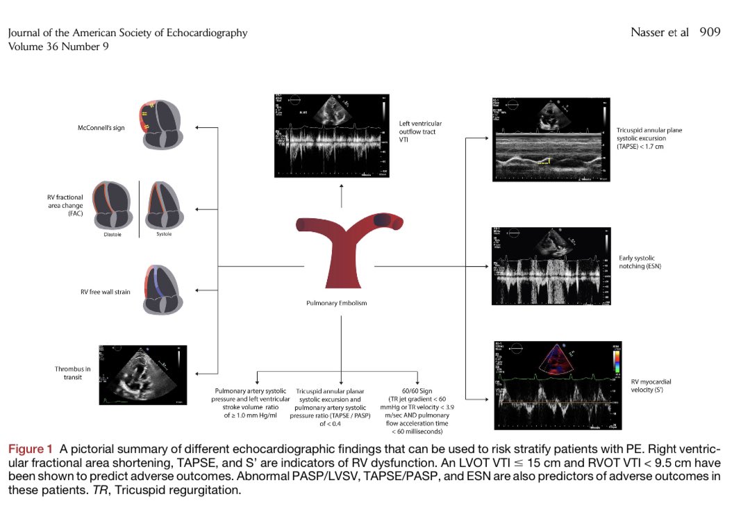
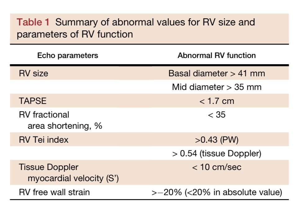
🫁 Evaluación ecocardiográfica de la embolia pulmonar: una revisión @ASE360 #CardioEd #Cardiology #echofirst #POCUS #ENARM

🫁 Evaluación ecocardiográfica de la embolia pulmonar: una revisión @ASE360 #CardioEd #Cardiology #echofirst #POCUS #ENARM

It's delightful to discuss #VExUS at the 3rd Nephro #criticalcare review course. Thanks to Dr. Tapas Kumar Sahoo and the team for extending the invitation and organizing this educational event. #Nephcrit #POCUS heralds the future!


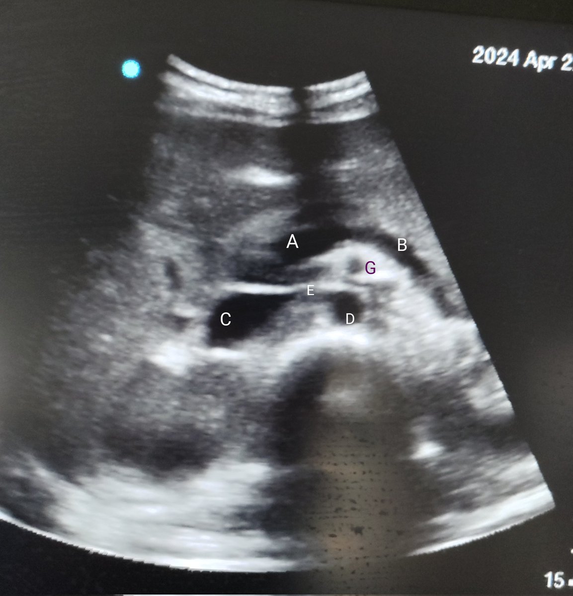
Abdominal ultrasound🫄 What are the labelled structures? #MedTwitter #MedX #FOAMED #POCUS #GITWITTER
@Rajiv_Sinanan 👏Physical exam (#POCUS) is powerful!

Amazing #POCUS session at #SCA2024! #Ultrasound techniques taught by THE BEST @scahq faculty!


@USaskUS @LindenKolbenson @TomGuzowski right back at @USaskUS @USUS_Regina ! Great work putting on a fun day of #POCUS learning with a good dose of hi-jinx! (👀 @ShawnSilver1989)

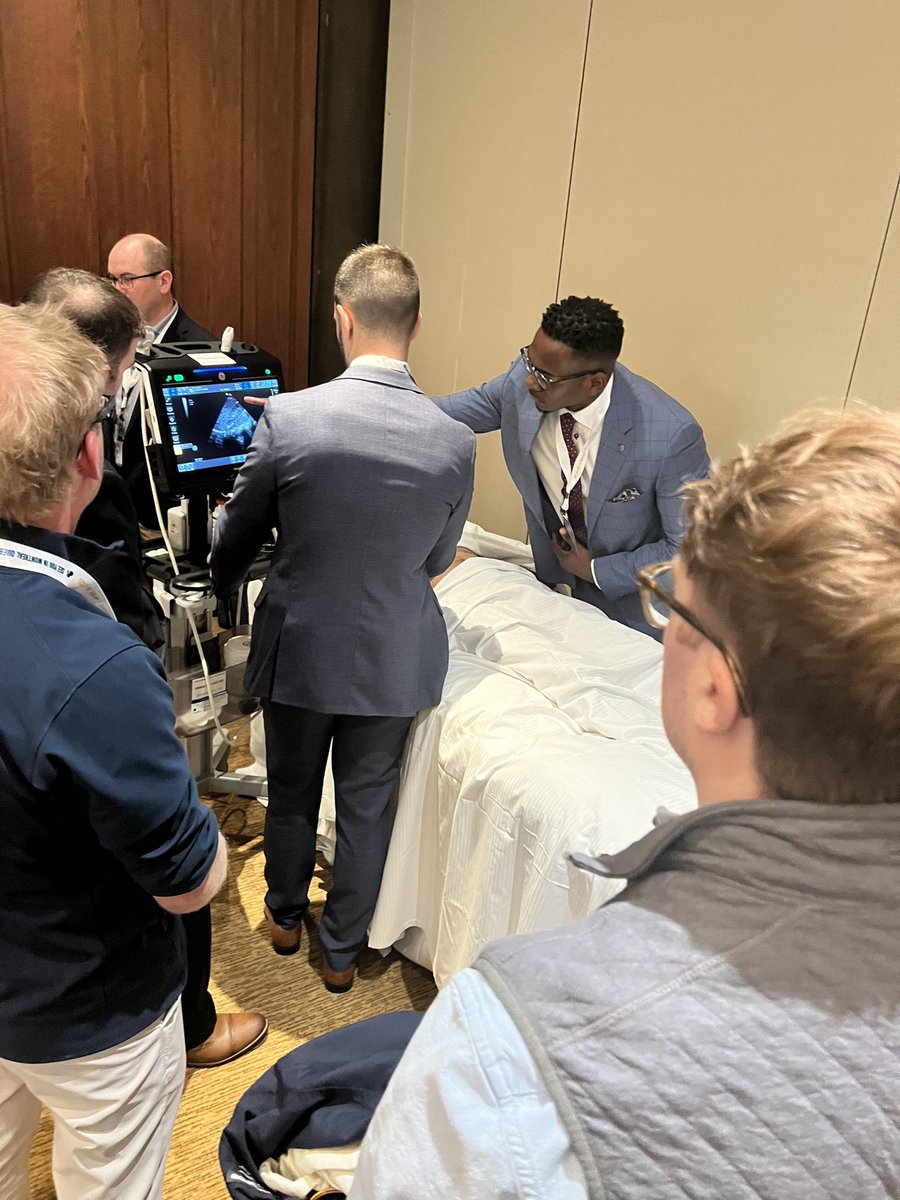
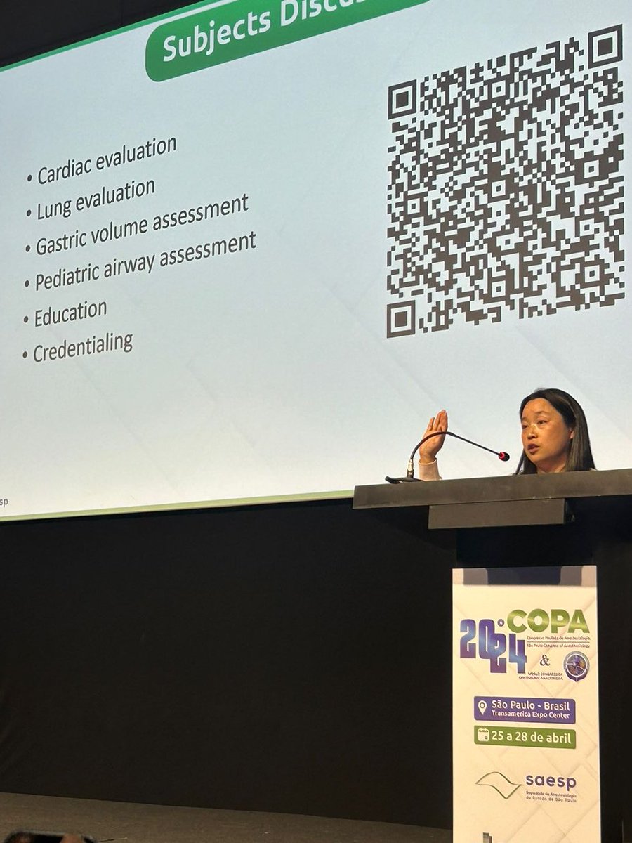
Thrilled to have @ElainaLin10 from @CHOP_GenAnesth lending her expertise in #PedsAnes #POCUS at #COPA24 @saesp! Huge thanks for coordinating our #POCUS Workshop and delivering an amazing lecture! @anestesia_usp @FMUSPoficial


Sometimes lungs can be decently anechoic making it decently difficult to decide if there is a pleural effusion. But if there are B lines coming from the diaphragm - that is lung. #POCUS #ultrasound #lungultrasound
New severe mitral regurgitation in patient with recent bacteremia is highly concerning for endocarditis. This was detected in less than a 5 minute bedside #pocus exam. #foamed #meded #medtwitter #cardiotwitter
Remarkable what can be done in minutes at the bedside with #pocus & @ButterflyNetInc IQ3 Elderly male with bacteremia treated 2 months prior presenting with progressive shortness of breath. JVD was not present in right IJ. Cardiac apical shown below:


PoCUS FOAMed @pocusfoamed
9K Followers 103 Following PoCUS FOAMed bring you the best free ultrasound education from around the web.

Practical POCUS @PracticalPOCUS
7K Followers 41 Following #POCUS #Training for #Hospitals, #EMS, #agencies, and #military while providing #FOAMed, #FOAMems, and #FOAMus information on #SoMe
POCUS 101 @Pocus101
16K Followers 47 Following Point of Care Ultrasound Made EASY for Anyone! #POCUS EM/ICU Doctor. Views are my own @viamdinh. RDMS, RDCS, and TEE (NBE) certified.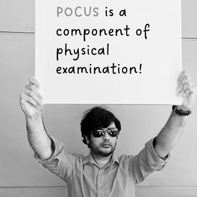
NephroPOCUS @NephroP
66K Followers 283 Following Point-of-care ultrasound #POCUS 📖 | by Abhilash Koratala MD @KoraAbhi, Associate Professor #Nephrology @MCW_Nephrology I👨🏻✈️ @POCUSIAPN | X≠ medical advice

POCUS La Paz @POCUSLaPaz
1K Followers 48 Following #POCUS teaching and research Institute at Hospital La Paz ED. Weekly Journal Club, cool cases and reviews! https://t.co/dj3MP74urc
THE POCUS Manifesto @POCUS_Manifesto
3K Followers 83 Following Expanding the Limits of the Physical Exam with Point-of-care Ultrasound. #pocus #laennecWouldPOCUS

Society of Point-of-C.. @POCUS_Society
14K Followers 1K Following Champion for POCUS-undertrained/underserved; non-profit; no industry COIs; #POCUS
CACTUS @POCUS_CACTUS
1K Followers 20 Following Children’s Acute Ultrasound (CACTUS) | UK #Paediatric #POCUS accreditation | in conjunction with Paediatric Intensive Care Society | Tweets by @MJGriksaitis
Hocus Pocus Tweets @HocusPocus1993
9K Followers 4 Following SISTAAAAAAAAHS! Hocus Pocus Appreciation Page ☆ (No Copyright Infringement Intended) ☆
IM POCUS Focus @IMPOCUSFocus
11K Followers 828 Following #IMPOCUS educational account sharing cases (fictionalized), ideas, EBM, and clinical integration. Not medical advice. Formerly PittIMPOCUS. #FOAMus.
Nicolas Lim @POCUSClub
4K Followers 200 Following 1st rule of POCUS club is 'you MUST talk about POCUS'. Ultrasound Leadership Academy Professor. Tweets about #POCUS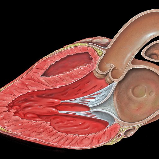
POCUS Echo @POCUSEcho
770 Followers 4 Following Collating Point-of-Care Critical Care Echo Images across the Twiitersphere

POCUS Med Ed @pocusmeded
11K Followers 249 Following Ultrasound vision for all. Must read: @pocus_manifesto
POCUS Certification A.. @POCUSAcademy
10K Followers 254 Following A non-profit offering #POCUS proficiency validation for all healthcare professionals. Part of https://t.co/ISvOzzNZZk. Tweets are not medical advice.
POCUS Bot @POCUSbot
1K Followers 0 Following Retweets of #POCUS or #FOAMus with minimum retweets of 5. Created by @DanMirsch. Sibling of @FOAM_bot
Hocus Pocus 2 | Now S.. @HocusPocusMovie
14K Followers 14 Following Hocus Pocus 2, an Original movie event, is now streaming on @DisneyPlus.
POCUS JEDI @JediPocus
2K Followers 844 Following Brazilian EM Doctors on Point-of-Care ultrasound education #POCUS #FOAMed #FOAMedBRA May the PROBE be with you! #POCUSJEDI
Pulmonary-POCUS @HoosierPocus
2K Followers 235 Following Pulmonary and Critical Care Medicine Point Of Care Ultrasound (POCUS) education. By @Edwin_J_Jr Website coming soon https://t.co/Yti0LE8HRQ
Hocus Pocus 2 | Now S.. @HocusPocus2US
2K Followers 88 Following Hocus Pocus 2, an Original movie event. Streaming September 30 only on @DisneyPlus .
Global Ultrasound Ins.. @GusiPocus
2K Followers 259 Following Global Ultrasound Institute (GUSI) is Transforming Patient Care Globally Through Online and In-Person Point-of-Care Ultrasound (#POCUS) Training & Education.
POCUS UK @POCUSUK
678 Followers 5 Following Uniting Knowledge & Skills in Point of Care Ultrasound (POCUS)| Democratising POCUS in a unique modern way| #FOAMed CoFounders @EveryOneNoOne1 & @NishCherian|
POCUS Journal @POCUSJournal
11K Followers 1K Following Journal of Point of Care Ultrasound | #FOAMus | #POCUS | #FOAMed | #POCUSJournal
The PoCUS Course @ThePoCUSCourse
785 Followers 16 Following The PoCUS Course - Pre-Hospital and Emergency Ultrasound Courses for Passionate Healthcare Professionals
NicuPocus @NicuPocus
3K Followers 183 Following Helping providers grow their neonatal point of care ultrasound knowledge and skills. Written by Alan Groves. All views his own. Disclaimer https://t.co/8EADMDn4wN
Alejandro @PleuralPOCUS
16K Followers 2K Following Respiratory Physician | Lung and Pleural POCUS | Chief of Pleural Diseases of @SMNCYT
Vancouver POCUS @VancouverPOCUS
887 Followers 67 Following Promoting multidisciplinary POCUS education in the lower mainland
The AIM POCUS Academy.. @AIM_POCUS
1K Followers 247 Following Founded August 2020 by Dr Andy Walden and Dr Joe Nunan to forward POCUS at the Royal Berkshire Hospital and beyond. #POCUS #FOAMEd #AIM
HEPOCUS (Liver POCUS) @hepocus
3K Followers 658 Following Hepatology related point- of-care Ultrasound. #POCUS. #HEPOCUS tweet Diego Arufe MD. Hepatologist. Hepatology and Liver Transplant. @GusiPocus Instructor
POCUS @POCUS_IG
807 Followers 18 Following Point of Care Ultrasound interest group at college of medicine, KSU @ksu_medicine , Part of @mscksu
CardioMotorizado @EmergPOCUS
7K Followers 2K Following Especialista en Medicina de Urgencias 🩻 | 📟 #POCUS Geek 💻 | #EchoFirst | Devotee of Emergency Ultrasound | #EchoViews 🫀| Reviews, Hilos y Posts.

Hocus Pocus 3 @HocusPocus3UK
2K Followers 77 Following They’re coming…..BACK!!! 🧹 #HocusPocus3 is officially in the works. 🕯️Contact Email: [email protected] ✨
Hocus Pocus Readathon @HPocusReadathon
2K Followers 325 Following Hello Witches! 🔮 Round 5: October 1st until 31st! 🎃 created by @deansgirlltiffy @nox_reads @larissasreading @fyrekatz1 @coffee_bookish
𝗗𝗮𝗻𝗶𝗲�.. @TaotePOCUS
4K Followers 1K Following 👶🏻Ⓜ️ | Taote - R2 Internal Medicine | Progressive Metal 🤘🎧 #POCUS
Temple Point-of-Care .. @TemplePOCUS
4K Followers 1K Following @TempleEM & @TempleIM Point-of-Care Ultrasound Program | Point-of-Care Ultrasound Education | Disclaimer: https://t.co/eUbUv6Fg8u | #POCUS #FOAMed #FOAMus
LocustPocus @CicadaLocPoc
611 Followers 203 Following Official page for 🪙 $CICADA 🪙 Community driven meme coin for the impending Cicada invasion. Get buzzing.
Hocus Pocus Studio & .. @hocuspocus_sjc
2K Followers 107 Following Desde 1994 promovendo a cultura independente! Rua Paraibuna, 838 - S. Dimas - São José dos Campos
HocusPocusMIA @HocusPocusMIA
1K Followers 39 Following Where princesses and ghouls meet. For one lovely purpose. To dance all night on their feet.
POCUS_Spain @POCUS_spain
2K Followers 107 Following @barbera_pablo👨🏻⚕️Urgenciólogo en el @HospitalLaFe (Valencia) Growing with POCUS 📲Instagram: POCUS_Spain 🎧: ECOCLINIC PODCAST
Saudi POCUS @SaudiPocus
334 Followers 13 Following Point of Care Ultrasound Workshop led by trained EMS Consultants. For inquiries: [email protected]
Joy Shen-Wagner @JOYofPOCUS
349 Followers 35 Following Family Medicine, POCUS Education, empowering primary care physicians, inspiring next generation of doctors, thoughts are my own.
Hocus Pocus @HocusPocusFi
4K Followers 2K Following Hocus Pocus is a DeFi protocol that leverages enchanting blockchain spells on the #PulseChain network | https://t.co/uPEub7locd
Hocus Pocus Edits! @HocusPocus_Edit
273 Followers 20 Following Lockscreen/wallpaper edit dedicated page for Anime, games and Kpop content! ✨Parcerias Purple Locks✨ pack by @purplelcks💜
pocus.sg @pocus_today
2K Followers 1K Following Dr Suresh Paranjothy, American Society of Anesthesiologists Local POCUS Mentor, SCCM Adult CCUS Instructor, Cardiac Anaesthetist, NUHS
Philips POCUS @PhilipsPOCUS
4K Followers 2K Following Bringing people & advanced technology together with a full suite of POCUS solutions. Breakthrough ultrasound tools for clarity in care delivery.
El Retorno de las Bru.. @HocusPocusSpain
1K Followers 7 Following Perfil en español de #HocusPocus (#ElRetornoDeLasBrujas). #HocusPocus3, ya confirmada #ElRetornoDeLasBrujas3
Hocus Pocus @hocpoc
15K Followers 166 Following Acoustic Hip Hop Band // 3 Albums, EPs & More Releases // 6 Members, including the Beatmaker/MC/DJ @mr20syl & @djgreem of @C2Cdjs // Label : @onandonrecords
Hocus Pocus: Vassar E.. @NuvancePOCUS
1K Followers 117 Following POCUS, EM: Our Advanced Clinical US program's goal is to deliver the finest possible training in the art and science of advanced clinical US.
P2|SK @ePOCUS
2K Followers 860 Following Improving the care of #injured and #ill children through the use of point-of-care #ultrasound (#POCUS) technology in #Toronto and abroad. #FOAMed #FOAMus
Global POCUS Partners @GlobalPocus
531 Followers 92 Following A consulting startup creating custom open-source hardware and software to facilitate remote point-of-care ultrasound (#POCUS) education anywhere in the world.
Alberto Gómez-Gonzá.. @FisioPocus
5K Followers 1K Following Physiotherapist on ICU 🇲🇽 |HGM🦅| Mechanical ventilation & Ultrasound #POCUS| #ECMO| Put your ICU in movement 🏥🏃♂️| I´m Straight Edge ✖️|
POCUS HUN-UNAL @HunPocus
963 Followers 221 Following Grupo de interés en Ultrasonido enfocado Hospital Universitario Nacional de Colombia y la Universidad Nacional. Canal Youtube: POCUS HUN

Hocus Pocus Guide @HocusPocusGuide
489 Followers 41 Following
Greymarch @CROnocusPocus
5K Followers 781 Following Mass adoption through mass education Tweeted, Twitted, Twoted Co-Founder of @PixelPalettePPN
Doom comes at dawn. �.. @MoreHocusPocus
1K Followers 253 Following Unholiness never appeared so attractive until it grew beneath my flesh. Alongside my sistren, we’re simply unstoppable. Beware. — parody.
Tanya Frane, RDMS, RM.. @TanyaPOCUS
1K Followers 926 Following #POCUS | Sonographer | Ultrasound Enthusiast
PoCUS East Point of C.. @PocusEast
222 Followers 46 Following PoCUS Training in Canada's Ocean Playground. Affiliated with the Canadian Point of Care Ultrasound Society. https://t.co/cJ9D4R0a7C #PoCUS #CPoCUS
Irene Ma @IM_POCUS
3K Followers 1K Following Professor of Medicine; Internal Medicine;#IMPOCUS; MD, MSc (Epi); PhD (MedEd), RDMS, RDCS, FRCPC, FACP, FAIUM 🏳️🌈 🇨🇦 gim_pocus on Threads
Scottish Neonatal Hae.. @ScotNeoPOCUS
701 Followers 190 Following All about neonatal cardiology, haemodynamics & point of care ultrasound including lung ultrasound!

Trading Pocus @tradingpocus
141 Followers 552 Following Curious about the market. #Gold #Silver #Copper #Uranium #Agri #Commodities $DJIA $SPX $TSX $VIX #USD #Cycles Tweets and RTs are not advice. Do your homework.
Pure Hocus Pocus @Body__Jewellery
790 Followers 0 Following Pure Hocus Pocus. Body Piercing, Body Jewellery, Alternative Clothing, Accessories. Based in King's Lynn, Norfolk, UK. Follow us on Facebook and Instagram.
Pediatric Global POCU.. @PocusGlobal
222 Followers 51 Following We are a collaboration of physicians dedicated to learning/teaching POCUS to help diagnose pediatric endemic diseases in resource limited settings.
RutgersNJMS_POCUS @RutgersPOCUS
1K Followers 322 Following Weekly @POCUSPearls — Rutgers Emergency Ultrasound Division @Rutgers_NJMS @RutgersNJMS_EM in Newark, NJ - @SAlerhand @annettemdinnyc @IOstrovsky99 @1indaqiu
POCUS Toronto @POCUS_Toronto
2K Followers 121 Following Point-of-Care Ultrasound @ University of Toronto. Ultrasound training from beginner to fellowship level.
Dr. Miguel Molina - E.. @dmiguelmolina
2K Followers 1K Following Docente de ecografía clínica Te ayudo a aprender ecografia clínica de manera sencilla

Locus Pocus @LocusKiwi
78 Followers 97 Following Rédacteur jeux vidéo, fanatique de Resident Evil 4, youtubeur à succès (relatif) (j'ai fais une vidéo) https://t.co/2KZib04Eux
Pocus @getpocus
637 Followers 124 Following The only Revenue Data Platform purpose-built for GTM. Analyze, visualize, and action data about your prospects and customers without needing engineers.


Hocus Pocus Studio @HocusPocusTweet
414 Followers 132 Following Hocus Pocus Studio is a leading London animation studio making films for brands, businesses and agencies across the world.
Paramedic Ultrasound @ParamedicPOCUS
757 Followers 934 Following Promoting advancement in #Paramedic practice through research & the implementation of #ParamedicUltrasound. Always looking to collaborate. #POCUS #foamUS
BC PoCUS @BCPoCUS
867 Followers 195 Following Your go-to resource for point-of-care ultrasound rapid summaries and videos. Brought to you by: Kevin Fairbairn Rural PoCUS enthusiast
NeonatalPOCUS @pocus_neonatal
935 Followers 337 Following Tweeting real-life neonatal POCUS images for education.
National Neonatal POC.. @NeoPocusCollab
573 Followers 521 Following National Collaborative ➡️ Expanding the use of #POCUS in the #NICU via universal curriculum, guidelines & credentialing ➡️ Advancing neo POCUS research & QI
Wokus pocus ✨🧙 @RhondaB42200045
971 Followers 780 Following Feisty Goth and loudmouth who won't keep quiet. Everything the Tories are not and the Gammon despise. Active mainly on Facebook. No DMs.#EU #fightfascism 🖖🇪🇺
Hocus Pocus Studio @HocusPocus_Lyon
533 Followers 221 Following Hocus Pocus is a Lyon based animation studio specialized in creation of CGI and visual effects for television, cinema and video game industry.
Hocus Pocus @hocuspocus_ar
2K Followers 956 Following
Focus Pocus @PupsherLive
1K Followers 2K Following Food/wine/jazz/books. #MLIS Creating a new groove. City spirit with small town roots. I ❤️animals, especially elephants.
Harefield POCUS @Harefield_POCUS
505 Followers 127 Following A multidisciplinary team at Harefield Hospital @ICU_Harefield delivering world-class #POCUS education. Stay tuned for information on our exciting new courses!
Okis @OkisPocus
848 Followers 415 Following retired pro fortnite pro and cod legend ||||| My Indiananimal @Dynamiiicc

DNF | HocusPocus35 @ipove_ssbu
1K Followers 228 Following @DNF_Esport smash ultimate, fgc player, SL & CRC
TheFrenchPOCUS @TheFrenchPOCUS
1K Followers 897 Following 🇫🇷 MD | Emergency medicine | Critical Care and Emergency Ultrasound #POCUS | Memesmith #VoltaireProtocol #NoFakeMed #FOAMed
MSK Ultrasound for PO.. @mskpocus
180 Followers 74 Following Quality, evidence based and clinically focused MSK Ultrasound training for Point-of-Care
The Opus Pocus @GrandmaDingley
404 Followers 839 Following Starring @BrianBlessed & @RoryBremner, 1001 Arabian Nights is the first tale from The Opus Pocus through which kids can discover the magic of classical musical!
PedsPOCUS @PedsPocus
352 Followers 175 Following For pediatric point-of-care ultrasound enthusiasts to connect with the community.

Manni D @OrizabaPocus
348 Followers 231 Following Organizer, e.r. doc commited to social justice and community health in México, #sinfronteras, #decolonization, autonomista, book lover and occasional poet
Pocus @Hrocusprocus
3K Followers 1K Following (T)aken. Just an acc to write what's on my mind. p.s. kalau belom di-followback, bilang aja ya hehe
Family Medicine POCUS.. @FMPOCUS_Comm
204 Followers 50 Following Fam Med Point-of-Care Ultrasound Interest Group Page | We aim to facilitate professional development and medical education pertaining to POCUS